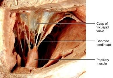
Structure From Which Chordae Tendineae Originate . Papillary muscle identify the features on this anterior view of the heart region of a cadaver by clicking and dragging the labels to the correct location. Structure from which chordae tendineae originate (h, papillary muscle) b.
Mitral Valve Chordae Tendineae. (A) Subvalvular Apparatus, View Of The... | Download Scientific Diagram from www.researchgate.net
Moreover, the fibers that constitute the chordae tendineae form bundles arranged in layers with a predominantly longitudinal disposition. After their origin and before their insertion, they split in numerous branches and interconnections that ensure a balanced distribution of the mechanical forces among chordae.
Mitral Valve Chordae Tendineae. (A) Subvalvular Apparatus, View Of The... | Download Scientific Diagram
They arise from the tips of the papillary muscles on the inside of the wall of the ventricles and extend into the hollow lumen. Prevents blood movement from right ventricle to right atrium. Structure from which chordae tendinae originate.
Source: www.knowyourbody.net
As observed with the scanning The other part was fixed in 2.5% morphogenesis of chordae tendineae in human [9] gluteraldehyde for transmission electrone microscopy lymphatic capillary in chordae [10] structure of chordae (tem. The chordae tendineae have their origin by the tip of the papillary muscles and insert on the rough zone of the mitral leaflets.
Source: slideplayer.com
They arise from the tips of the papillary muscles on the inside of the wall of the ventricles and extend into the hollow lumen. Structure from which chordae tendinae originate winters and puny line | wurzel hasnicards by nweismanuy bio tou The tricuspid valve moves up and closes the opening between the right atrium and right ventricle.
Source: www.coursehero.com
The chordae tendineae make up the leaflet suspension system that ultimately determine and maintain the position and tension on the valve leaflets at end of systole. Pulmonary valve has three cusps that open when the right ventricle contracts. Although the anatomy and function
Source: www.researchgate.net
The chordae originate from the fibrous heads of the papillary muscles and may be classified according to their site of insertion on the leaflet. Prevents blood movement from right ventricle to right atrium. Prevents blood movement from the right ventricle to right atrium tricuspid valve.
Source: study.com
Are part of the subvalvular apparatus, the chordae tendinae are composed of fibrous strings that originate from the papillary muscles or the ventricle wall and that its insertion is into the ventricle, the anterior leaflet, posterior leaflet, and commissural leaflet. The chordae tendineae have their origin by the tip of the papillary muscles and insert on the rough zone of.
Source: www.chegg.com
The tricuspid valve is made up of 3 large cusps with chordae tendineae while the pulmonary valve is made up of 3 small cusps without chordae tendineae. Pulmonary valve has three cusps that open when the right ventricle contracts. The tricuspid valve moves up and closes the opening between the right atrium and right ventricle.
Source: www.chegg.com
The chordae originate from the fibrous heads of the papillary muscles and may be classified according to their site of insertion on the leaflet. The mitral valve (mv) is a very complex structure composed of the anterior (aml) and posterior (pml) leaflets, the chordae tendineae (ct), and the papillary muscles (ppmm), all of which work on a very harmonious basis.
Source: www.researchgate.net
The chordae tendineae have their origin by the tip of the papillary muscles and insert on the rough zone of the mitral leaflets. A single papillary muscle may originate multiple chordae tendineae which can divide into branches in swine and human hearts. Where connective tis sue originate
Source: www.chegg.com
Are part of the subvalvular apparatus, the chordae tendinae are composed of fibrous strings that originate from the papillary muscles or the ventricle wall and that its insertion is into the ventricle, the anterior leaflet, posterior leaflet, and commissural leaflet. They have central function by allowing coaptation and preventing leaflet prolapse or flail into the left atrium. Structure from which.
Source: www.pharmacy180.com
Multiple chordae tendineae attach to each leaflet of each flap of the valves. Membranes around the heart pericardial sac. Moreover, the fibers that constitute the chordae tendineae form bundles arranged in layers with a predominantly longitudinal disposition.
Source: www.researchgate.net
The mitral valve (mv) is a very complex structure composed of the anterior (aml) and posterior (pml) leaflets, the chordae tendineae (ct), and the papillary muscles (ppmm), all of which work on a very harmonious basis to ensure an appropriate opening and closing of the left atrioventricular orifice. Are part of the subvalvular apparatus, the chordae tendinae are composed of.
Source: studylib.net
Prevents blood movement from left ventricle to left atrium mitral valve. Membranes around heart (j, pericardial sac) d. Multiple chordae tendineae attach to each leaflet of each flap of the valves.
Source: www.knowyourbody.net
Pulmonary valve has three cusps that open when the right ventricle contracts. False chordae tendineae are irregular and more commonly are attached either between papillary muscles or from the papillary muscles to the walls of the ventricles. Papillary muscle identify the features on this anterior view of the heart region of a cadaver by clicking and dragging the labels to.
Source: socratic.org
Moreover, the fibers that constitute the chordae tendineae form bundles arranged in layers with a predominantly longitudinal disposition. The tricuspid valve is large and has chordae tendinaea and papillary muslces. Structure from which chordae tendinae originate.
Source: www.chegg.com
False chordae tendineae are irregular and more commonly are attached either between papillary muscles or from the papillary muscles to the walls of the ventricles. The chordae tendineae have their origin by the tip of the papillary muscles and insert on the rough zone of the mitral leaflets. Describe the function of the chordae tendineae and the papillary muscles.
Source: studylib.net
As observed with the scanning The chordae originate from the fibrous heads of the papillary muscles and may be classified according to their site of insertion on the leaflet. After their origin and before their insertion, they split in numerous branches and interconnections that ensure a balanced distribution of the mechanical forces among chordae.
Source: en.wikipedia.org
The simplest and perhaps most effective classification of chordae tendineae. Chordae tendineae contain elastin in. A single papillary muscle in swine and human hearts may originate several chordae tendineae which after that may divide in several branches as described previously (lam et al.;
Source: www.heartviews.org
41,46 after their origin and before their insertion, they split in numerous branches and interconnections that ensure a balanced distribution of the mechanical forces among chordae. As observed with the scanning Pulmonary trunk_____ gives rise to left and right pulmonary arteries
Source: clinicalanatomy.com
False chordae tendineae are irregular and more commonly are attached either between papillary muscles or from the papillary muscles to the walls of the ventricles. Structure from which chordae tendineae originate papillary muscle. The tricuspid valve moves up and closes the opening between the right atrium and right ventricle.
Source: www.researchgate.net
They have central function by allowing coaptation and preventing leaflet prolapse or flail into the left atrium. Chordae tendineae contain elastin in. These issues originate from papillary muscles.