Which Type Of Microscope Shows Cells Against A White Background . Darkfield microscope formed a bright image against dark background: The macrophage cells are an essential component of the immune system, which is the body’s defense against potential pathogens and degraded host cells.
183,561 Microscope Stock Photos, Pictures & Royalty-Free Images - Istock from www.istockphoto.com
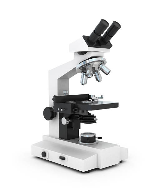
The common light microscope used in the laboratory is called a compound microscope because it contains two types of lenses that function to magnify an object. To increase contrast, the technician inserts an opaque light stop above the illuminator.
183,561 Microscope Stock Photos, Pictures & Royalty-Free Images - Istock
Compound microscopes are light illuminated. 03.07 list and describe the three elements of good microscopy. This microscope shows cells against a bright background and also shows intracellular structures of unstained cells based on their varying densities:
Source: www.coursehero.com
Compound microscopes are light illuminated. Transmission em is used for internal detail of cells and subcellular structures. This microscope is the most widely used and shows cells against a bright background.
Source: www.coursehero.com
Male hand in blue protective gloves holding. The macrophage cells are an essential component of the immune system, which is the body’s defense against potential pathogens and degraded host cells. The common light microscope used in the laboratory is called a compound microscope because it contains two types of lenses that function to magnify an object.
Source: www.coursehero.com
The image seen with this type of microscope is two dimensional. 4 shows the image sample after color space. Surface of the red blood cells and the antibodies are in the.
Source: opentextbc.ca
What microscope shows cells against a bright background and also shows intracellular structures of unstained cells based on their varying densities? Male hand in blue protective gloves holding. The macrophage cells are an essential component of the immune system, which is the body’s defense against potential pathogens and degraded host cells.
Source: www.nature.com
The lens closest to the eye is called the ocular, while the lens closest to the object is called the objective. 03.07 list and describe the three elements of good microscopy. That’s the major difference between plant and animal cells under microscope.
Source: www.chegg.com
This microscope is the most widely used and shows cells against a bright background. The common light microscope used in the laboratory is called a compound microscope because it contains two types of lenses that function to magnify an object. Most microscopes have on their base an apparatus called a condenser, which condenses light.
Source: www.technologynetworks.com
Comparing transmission electron microscopy with scanning electron microscopy, the following statement is true. You can view individual cells, even living ones. Under the brightfield microscope, the technician can barely see the bacteria cells because they are nearly transparent against the bright background.
Source: www.gettyimages.com
Which type of microscope is the most widely used and shows cells against a bright background? The bending of light rays as they pass from one medium to another is called refraction. Generalized cell is used for structure of animal cell and plant cell to present the.
Source: www.coursehero.com
Which type of microscope shows cells against a white background? You know, animal cell structure contains only 11 parts out of the 13 parts you saw in the plant cell diagram, because chloroplast and cell wall are available only in a plant cell. This microscope is the most widely used and shows cells against a bright background.
Source: www.chegg.com
This microscope shows cells against a bright background and also shows intracellular structures of unstained cells based on their varying densities: Transmission electron microscope (tem) description. Bright field dark field phase contrast fluorescence electron
Source: www.gettyimages.com
Which type of microscope shows cells against a bright background and also shows intracellular structures of unstained cells based on their varying densities? Male hand in blue protective gloves holding test tube with blood sample against background of microscope. Surface of the red blood cells and the antibodies are in the.
Source: www.coursehero.com
Consequently, the cell appears as a bright object against a dark background. Which type of microscope is the most widely used and shows cells against a bright background? Generalized cell is used for structure of animal cell and plant cell to present the.
Source: www.nicepng.com
Which type of microscope shows cells against white background? Which type of microscope shows cells against a bright background but also differentiates intracellular structures of unstained cells based on their varying densities? Under the brightfield microscope, the technician can barely see the bacteria cells because they are nearly transparent against the bright background.
Source: www.coursehero.com
Most microscopes have on their base an apparatus called a condenser, which condenses light. A) bright fieldb) dark fieldc) phase contrastd) differential interferencee) electron. The bending of light rays as they pass from one medium to another is called refraction.
Source: www.coursehero.com
This microscope shows cells against a bright background and also shows intracellular structures of unstained cells based on their varying densities. Which type of microscope shows cells against a white background? Which type of microscope shows cells against a bright background but also differentiates intracellular structures of unstained cells based on their varying densities?
Source: www.coursehero.com
Consequently, the cell appears as a bright object against a dark background. Transmission electron microscope (tem) description. Under the brightfield microscope, the technician can barely see the bacteria cells because they are nearly transparent against the bright background.
Source: www.chegg.com
This microscope shows cells against a bright background and also shows intracellular structures of unstained cells based on their varying densities. 03.07 list and describe the three elements of good microscopy. A simpler way to see some of the features of a living cell is to observe the light that is scattered by its various components.
Source: www.coursehero.com
Bright field dark field phase contrast fluorescence electron Which type of microscope shows cells against a bright background but also differentiates. Darkfield microscopes have a device to scatter light from the illuminator so that the specimen appears white against a black background.
Source: www.gettyimages.com
This microscope shows cells against a bright background and also shows intracellular structures of unstained cells based on their varying densities. Transmission em is used for internal detail of cells and subcellular structures. Which type of microscope shows cells against white background?
Source: www.istockphoto.com
This microscope achieves the greatest resolution and highes magnification. The macrophage cell is a large cell derived from a monocyte, a type of blood cell, which enters the connective tissue matrix from the blood vessels. You know, animal cell structure contains only 11 parts out of the 13 parts you saw in the plant cell diagram, because chloroplast and cell.